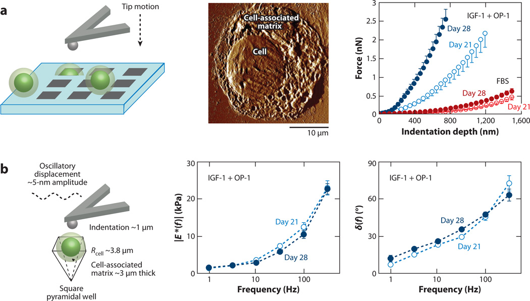
Figure 4.
Temporal development of tissue-engineered, chondrocyte-associated matrix. (a) (Left) Schematic of indentation (using a spherical tip R ~ 2.5 µm) on individual chondrocytes and their cell-associated matrix, which are fixed within pyramidal wells of a silicon microfabricated substrate. (Middle) Tapping-mode atomic force microscopy image of a chondrocyte and newly synthesized tissue-engineered cell-associated matrix after 11-day culture in 10% fetal bovine serum (FBS). (Right) Nanoindentation curve (mean ± SEM of five loading cycles per cell for n ≥ 5 cells) on loading of individual chondrocytes with engineered cell-associated matrix (after release from alginate at different times) in culture with 10% FBS and with insulin-like growth factor-1 (IGF-1) and osteogenic protein-1 (OP-1). Panel a adapted with permission from Reference 11. (b) Dynamic mechanical properties of chondrocytes and their engineered cell-associated matrix (mean ± SEM; n = 5 cells) using a spherical tip in culture with IGF-1 and OP-1. (Left) Schematic of the dynamic oscillatory nanoindentation. (Middle) Dynamic indentation modulus, |E*|, and (Right) the phase angle, δ, as a function of oscillation frequency after 21 and 28 days of culture. Panel b adapted with permission from Reference 12.

