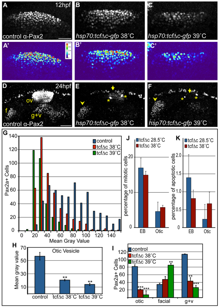
Fig. 7.

Inhibition of Wnt signaling reduces Pax2a levels resulting in a cell segregation shift from otic to facial placode. (A-C′) Confocal projections from control and Tg(hsp70:tcfΔC-EGFP) zebrafish embryos heat-shocked at 10 hpf and analyzed for Pax2a expression at 12 hpf. Note decreased Pax2a levels in heat map images as heat-shock stringency increases (A′-C′). (D-F) Control and Tg(hsp70:tcfΔC-EGFP)+ embryos heat-shocked at 10 hpf and analyzed for Pax2a expression at 24hpf (arrowheads, facial placode; arrows, otic vesicle; asterisks, putative otic sensory patches). (G) PPA Pax2a expression MGVs at 12 hpf. Note significant shift from high to low Pax2a levels with increased heat-shock stringency (χ2-test; P<<0.001). (H) Average MGV of Pax2a expression in the otic vesicle was significantly reduced in Tg(hsp70:tcfΔC-EGFP)+ embryos (heat-shocked at 39°C) versus controls (n≥317 cells from five embryos per condition; unpaired t-test; **P<0.003). (I) Quantification of Pax2a+ cells in control and Wnt-inhibited embryos (heat-shocked at 39°C) reveals significant otic vesicle and glossopharyngeal/vagal placode cell losses, with concurrent increases in facial placode (***P<0.001; **P<0.01). (J) Percentage of mitotic cells (by pH3 immunolabeling) at 18 hpf following heat-shock of Tg(hsp70:tcfΔC-EGFP) embryos at 10 hpf was unchanged versus uninduced controls (n=12 embryos per condition). (K) There is no significant change in Caspase-3+ cell percentage at 18 hpf following heat-shock of Tg(hsp70:tcfΔC-EGFP) embryos at 10 hpf (n=12 embryos per condition). Error bars represent s.e.m. f, facial placode; g+v, glossopharyngeal/vagal placodes; MGV, mean gray value; ov, otic vesicle. Scale bars: 50 μm.
