
Fig. 3.
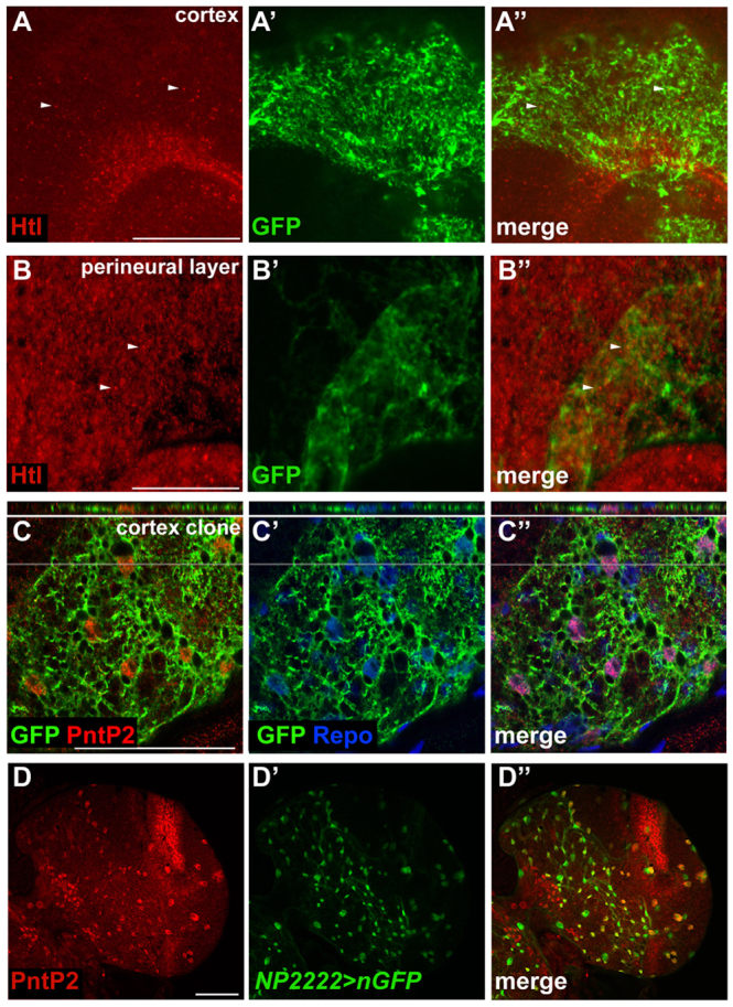
Expression of FGF pathway components in the larval brain. (A-B″) Htl expression (red) in glia in the cortex (A-A″) or perineural (B-B″) layers of the Drosophila larval brain. repo-MARCM clones expressing GFP (green) were used to identify the respective glial types. (C-C″) Third instar larval brain hemisphere showing a repo-MARCM cortex clone stained for GFP (green), PntP2 (red) and Repo (blue) expression. Note that PntP2-positive cortex glia colocalise with GFP in the orthogonal view (top; grey lines indicate the position of orthogonal section). (D-D″) Cortex glia-specific NP2222-Gal4 driving UAS-nGFP (green) showing colocalisation with PntP2 expression (red). Scale bars: 50 μm.