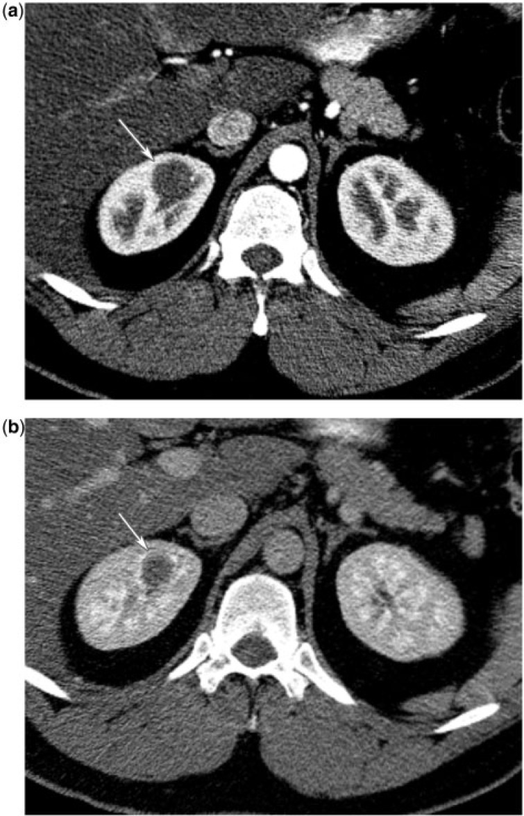
Figure 16.
(a) A 35-year-old man with right renal hemangioma. CT renal protocol in the corticomedullary phase reveals a poorly enhancing mass in the upper pole medulla of the right kidney (arrow). (b) A 35-year-old man with a right renal hemangioma. CT renal protocol in the nephrographic phase reveals a poorly enhancing mass in the upper pole medulla of the right kidney (arrow).

