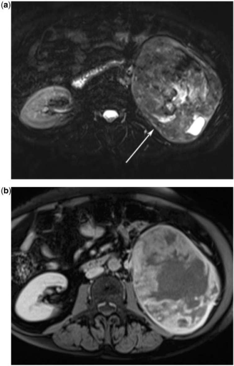
Figure 19.
(a) A 71-year-old woman with renal leiomyosarcoma. T-2 weighted fat-saturates axial MR image reveals a mixed signal, well-circumscribed mass replacing the left kidney (arrow). (b) A 71-year-old woman with renal leiomyosarcoma. T1-weighted fat-saturated post intravenous contrast axial MR image shows that the mass has heterogeneous enhancement.

