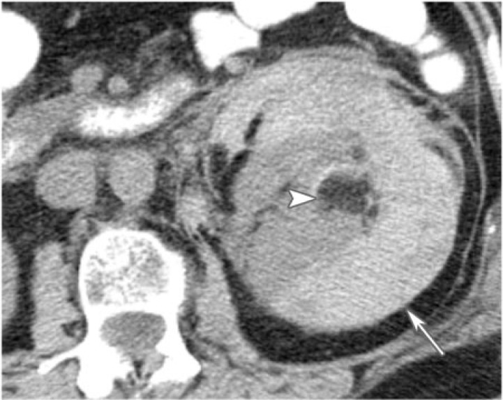
Figure 6.
A 74-year-old woman with left perinephric hematoma from a bleeding angiomyolipoma. CT without intravenous contrast shows a high attenuation fluid collection surrounding the left kidney. The high attenuation fluid collection is an acute hematoma (arrow). The fat attenuation mass is the bleeding angiomyolipoma (arrowhead).

