Figure 3.
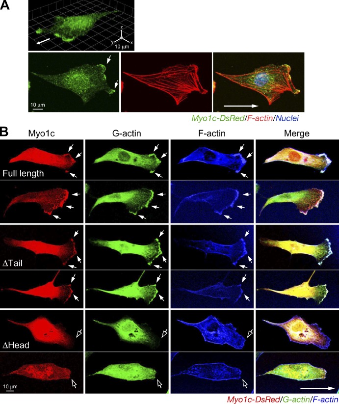
Motor domain–dependent localization of Myo1c at the cell leading edge. (A) Myo1c localization at the cell leading edge. ECs were stained with anti-Myo1c antibody and visualized with Alexa Fluor 488–IgG and Alexa Fluor 568–phalloidin. 3D reconvolution (top) and single-layer confocal scanning images are shown. Arrows indicate the direction of cell migration. (B) Localization of Myo1c at the leading edge requires the motor domain. ECs were transfected with plasmids encoding full-length or domain-deleted Myo1c fused with DsRed. Migrating cells were stained with Alexa Fluor 488–DNase I. Filled arrows indicate colocalized Myo1c and G-actin at the leading edge, and open arrows show the leading edge with minimal colocalization.

