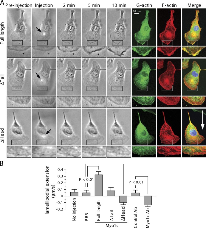
Figure 4.
Myo1c-induced actin dynamics in lamellipodia. (A) Representative images. Microinjection of Myo1c induces rapid G-actin accumulation and plasma membrane ruffling at the leading edge, increasing lamellipodial extension. Cells were microinjected with purified full-length or truncated Myo1c, and cell morphology was monitored for 10 min followed by staining for visualization of G- or F-actin. Arrows indicate the microinjection spots. The boxed areas are magnified below each image. The white arrow indicates the direction of cell migration. (B) Quantification of lamellipodial extension/retraction speed (mean ± SEM; n = 5–9 cells). Ab, antibody.

