Figure 2.
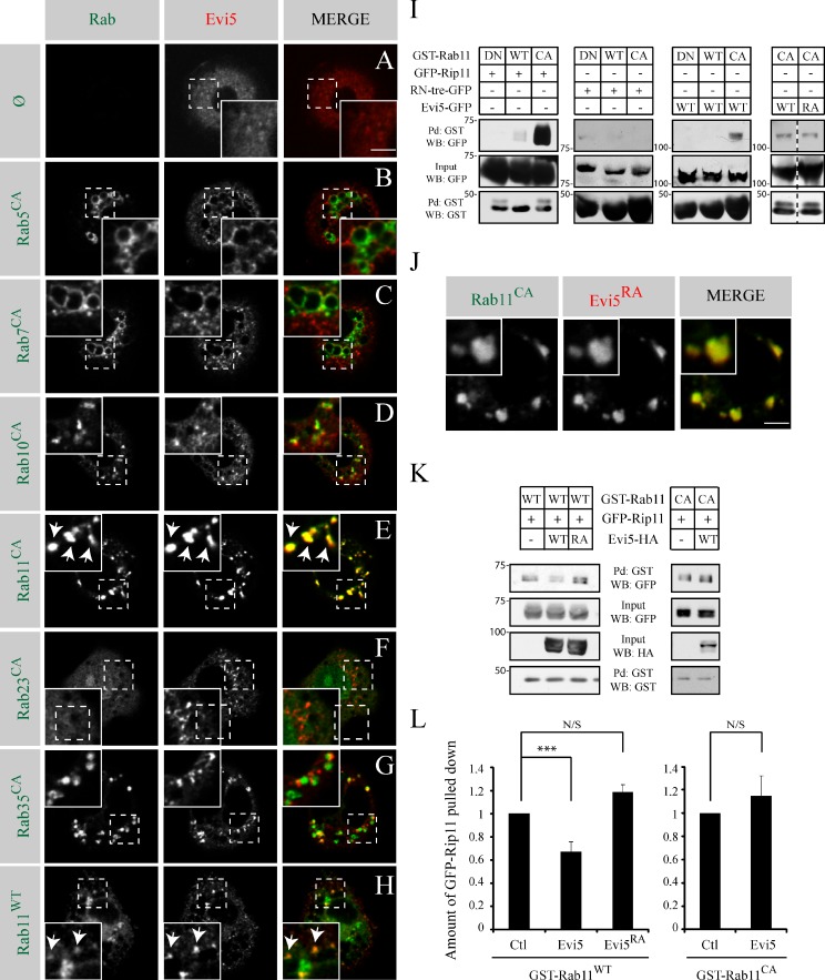
Evi5 acts as a Rab11-GAP in vitro. (A–H) Representative images of S2 cells transfected with Evi5-mcherry and the indicated GFP- or YFP- tagged form of Rab. Rab proteins are either expressed in their wild-type (WT) or CA form. A grayscale image of both the green and red channel is shown for every image. The insets show higher magnifications of the regions marked by dashed line squares. Arrows point to structures where Evi5 and Rab11 colocalize. (I) S2 cells were cotransfected with the wild-type, DN, or CA forms of GST-Rab11 together with the indicated GFP constructs. Pull-downs (Pd) using glutathione beads were performed on lysates. Proteins bound to GST-Rab11 were detected by Western blotting (WB). (J) YFP-Rab11CA was coexpressed with Evi5RA-mcherry in S2 cells. Insets show higher magnification. (K) Effector pull-down assays performed with lysates from S2 cells cotransfected with GST-Rab11WT (left) or GST-Rab11CA (right) together with GFP-Rip11 with or without HA-Evi5. Pull-down of GST-Rab11 and protein analysis were performed as in I. (L) Quantification of the total pulled down GFP-Rip11 normalized to the control on three (GST-Rab11CA) or four (GST-Rab11WT) independent experiments. (***, P < 0.05; t test). Ctl, control. Error bars are standard error of the mean. Bars, 5 µm.

