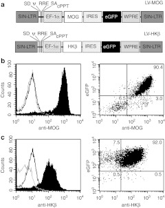
Figure 1.
Schematic representation of recombinant lentiviral vectors and in vitro expression of engineered transgenes. (a) The proviral, self-inactivating (SIN) forms of LV-MOG or LV-HKβ lentiviral vectors encoding mouse MOG or the β subunit of the hydrogen-potassium cDNA, respectively, under the control of the EF-1α promoter. Both vectors incorporate an eGFP tag under the translational control of an internal ribosomal entry site (IRES) sequence. (b) Expression of MOG or (c) HKβ in transduced 427.1 cells as determined by flow cytometry. Left hand panel shows expression profile of cells transduced with LV-MOG or LV-HKβ and stained with anti-MOG or anti-HKβ antibodies, respectively (black shaded curve). Isotype staining (grey curve) and staining of transduced 427.1 cells expressing an irrelevant antigen (black curve) served as controls. Dot plot analysis of trasduced 427.1 cells co-expressing eGFP and MOG or HKβ (right hand panel). Proportions of MOG:eGFP and HKβ:eGFP subsets are numerically indicated in each quandrant. Data are representative of two independent experiments. EF, elongation factor; eGFP, enhanced green fluorescent protein; LTR, long-term repeat; LV, lentiviral vector; MOG, myelin oligodendrocyte glycoprotein; ψ, packaging signal; RRE, rev response element; SA, splice acceptor site; SD, splice donor site; WPRE, woodchuck hepatitis virus post-transcriptional regulatory element.

