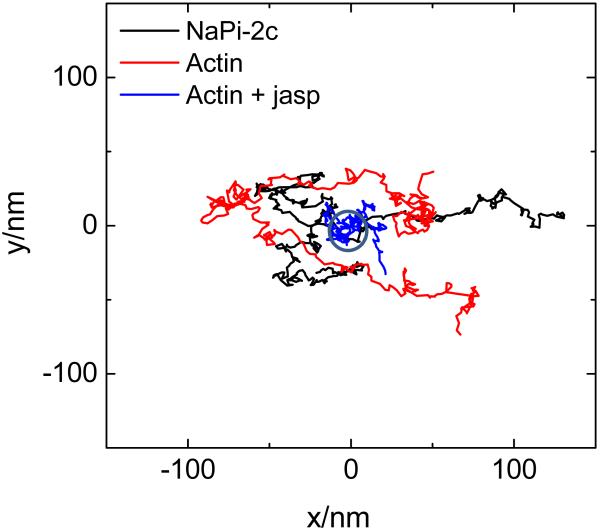
Figure 4.
Dynamics of the center of mass of microvilli. The z position of the scanner was kept constant while the feedback mechanism followed the motion of the center of mass of the microvilli. Each measurement is done different microvilli according to the legend color. Two different proteins were imaged, Cerulean NaPi 2C which resides on the microvilli surface and Actin EGFP which presumably is in the cytoplasm. Stabilization of actin polymerization was obtained by treatment with Jasplakinolide.

