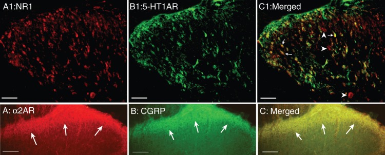
Fig 2.
(a1–c1) Immunostaining microphotographs show that 5-HT1ARs are located in NR-containing neurones in the spinal dorsal horn. Arrows point to double-stained neurones; arrow heads point to NR1-containing neurones. Scale is 50 μm. (a–c) Immunostaining microphotographs show that α2a-ARs are located in CGRP-containing primary afferents in the spinal cord. Arrows point to superficial laminae. Scale is 100 μm.

