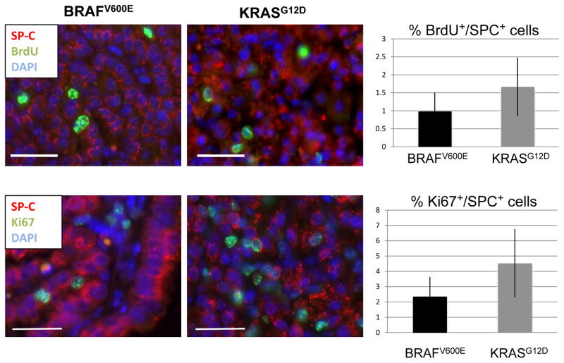
Figure 2. Quantification of proliferating cells in BRAFV600E and KRASG12D driven lung tumors.
A: BrdU (green) and SP-C (red) staining of BRAFV600E and KRASG12D driven tumors at 17 weeks. Double positive cells within fully formed adenomas were classified as proliferating. Bar represents 25μm. Images are 40× magnification.
B: Ki67 (green) and SP-C (red) staining of BRAFV600E and KRASG12D driven tumors at 17 weeks. Bar represents 25 μm. Images are 40× magnification.

