
Figure 8. T cell response to the capsid by different AAV serotypes.
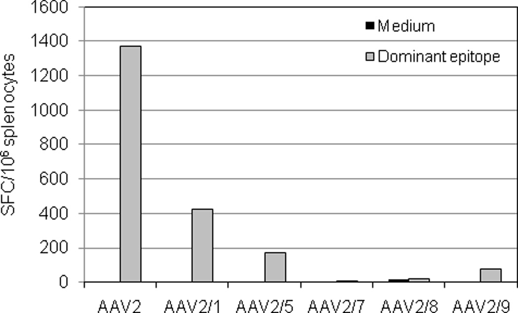
BALB/c mice were intramuscularly injected with AAV vectors at 1×10¹² GC/mouse (n=3 per vector). Seven days after injection, splenocytes were isolated, pooled, and stimulated with H-2d-restricted dominant epitopes (AAV2: VPQYGYLTL; other serotypes: IPQYGYLTL) or medium alone for IFN-γ ELISPOT assay. Peptide-specific cells are presented as spot forming cells (SFC) per 106 splenocytes.