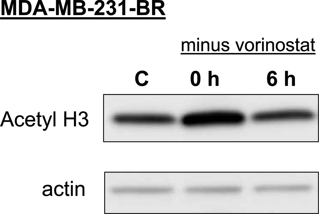
Figure 1.
Histone acetylation status determined after exposure to and removal of vorinostat. MDA-MB-231-BR cells were either exposed to vorinostat (1 µmol/L) for 16 h and collected (time 0 h) for immunoblot analysis of acetylated histone H3 or exposed to vorinostat (1 µmol/L) for 16 h; medium was removed; and cells were rinsed with PBS, fed fresh drug-free medium, and collected 6 h later for immunoblot analysis. C, cultures exposed to the vehicle only (DMSO).

