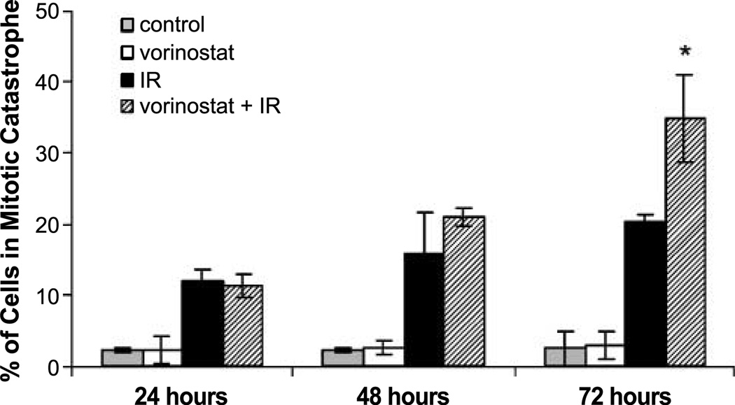
Figure 4.
Mitotic catastrophe results of MDA-MB-231-BR cells. Gray columns, vehicle-treated cells; white columns, cells treated with vorinostat (1 µmol/L) alone; black columns, cells treated with irradiation alone (2 Gy); hatched columns, cells treated with the combination of vorinostat (1 µmol/L) and irradiation (2 Gy). Vorinostat was given 16 h before treatment and maintained in the medium until cells were collected at designated time points. Columns, mean; bars, SE. Nuclear fragmentation was defined as the presence of two or more distinct lobes within a single cell. *, P < 0.01, according to Student's t test (irradiation versus vorinostat + irradiation).

