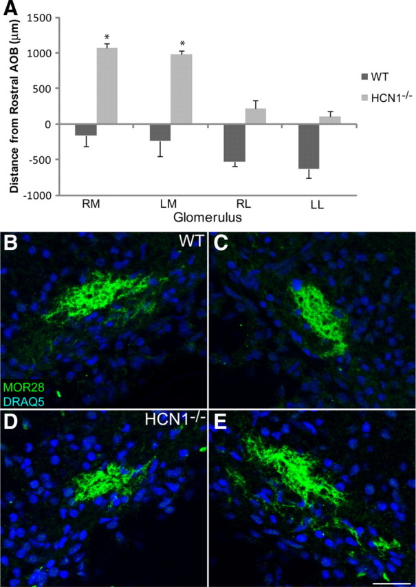
Figure 11.

In HCN1−/− mice MOR28 glomeruli change their location but have normal coalescence. A, The histogram shows the change in position relative to the first section containing AOB in the WT compared to HCN1−/− mice. B–E, OB sections from P0 mice labeled with MOR28 and DRAQ5. B, C, Left and right medial WT glomeruli, respectively. D, E, Left and right medial HCN1−/− glomeruli, respectively. RM, Right medial; LM, left medial; RL, right lateral; LL, left lateral. Scale bar for B–E (in E), 50 μm. Error bars, SEM. *p < 0.05, statistically significant difference from controls.
