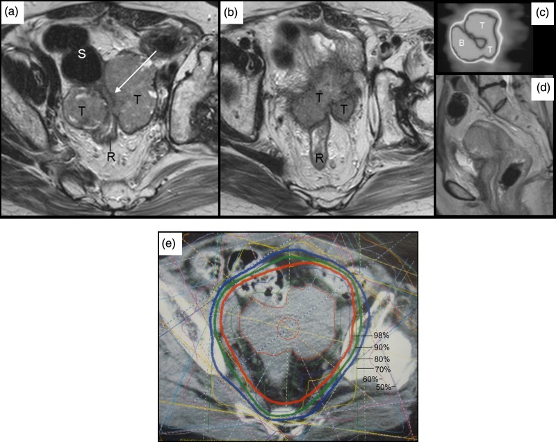
Fig. 3.
MR images before external beam treatment show recurrent tumor (T) tightly encasing a part of the rectosigmoidal colon (long white arrow) (a, b). Sagittal image of FDG-PET study showing increased accumulation at maximum standardized uptake value of 4.56 (confirming) (c) and a corresponding MR image (d). Dose distributions on X-ray CT image in conformal radiotherapy.

