Fig. 2.
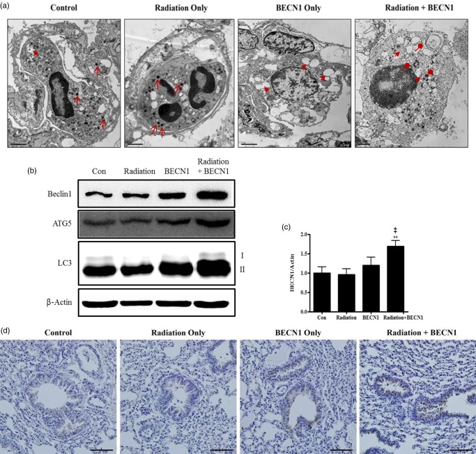
Delivery of beclin1 via inhalation was successful and activated the autophagy pathway.
(a) Intracellular lung structures were screened under transmission electron microscopy. Arrows, primary lysosomes; arrow-heads, cytoplasmic vacuoles; circle-heads, secondary lysosomes/autophagolysosomes. Magnification: ×6000. Scale bar: 1 µm. (b) Western blot was performed to monitor the increase in autophagy-related proteins: beclin1 (1:500 dilution), ATG5 (1:1000 dilution) and LC3 (1:1000 dilution). Bands are representative of five individuals from each group. (c) Densitometric analyses reconfirmed the synergistic effect of beclin1 on autophagy. Each bar represents the mean ± SE (n = 5). **P < 0.05 was considered highly significant compared with the control group and ‡P < 0.01 was highly significant compared with the radiation group. (d) Delivery of beclin1 and its synergistic effect in combination group were confirmed with IHC analysis; Beclin1 (1:100 dilution). Magnification: ×200. Scale bar: 20 µm. Representative figures of five mice per group.

