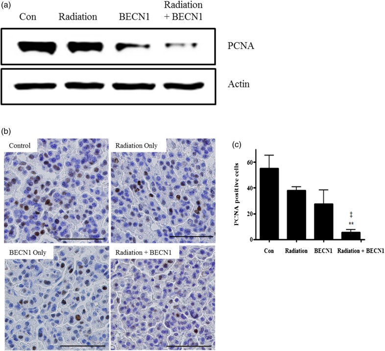
Fig. 4.
Beclin1 with radiation decreased cell proliferation in lungs of K-rasLA1 mice.
(a) Western blot of anti-PCNA (1:5000 dilution) and the lung tissue lysates. Bands are representative of five individuals from each group. (b) The immunohistochemistry analysis showed fewer double stained nuclei with hematoxylin and chromogen on lung tissue slides. Magnification: ×400. Scale bar: 50 µm. (c) Statistical analysis of PCNA positive cells. Nuclei double-stained with DAB and hematoxylin were counted in three different fields from each slide. Each bar represents the mean ± SE (n = 5). **P < 0.01 was considered highly significant compared with the control group. ‡P < 0.01 was considered highly significant compared with the radiation group. Representative figures of five mice per group.

