
Fig 2.
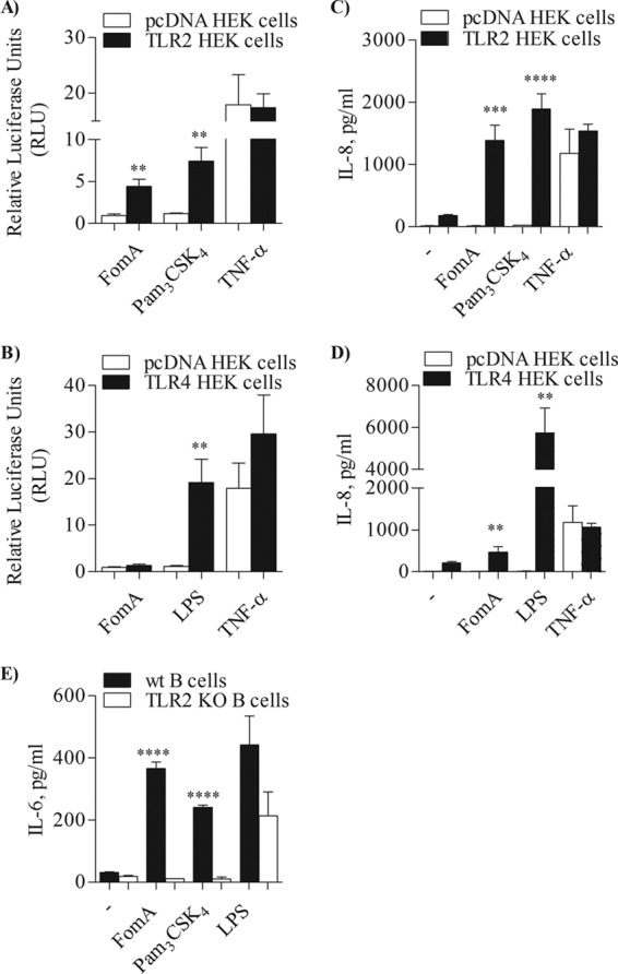
TLR2-dependent cell activation by FomA. (A and B) TLR2-HEK cells and pcDNA-HEK cells (A) or TLR4-HEK cells and pcDNA-HEK cells (B) incubated with purified FomA (10 μg/ml), Pam3CSK4 (100 ng/ml), E. coli LPS (100 ng/ml), and TNF-α (20 ng/ml) for 18 h. NF-κB-dependent luciferase activity was measured and is expressed in arbitrary units ± SD normalized to nonstimulated cells. **, P < 0.0023 (for panel A) and P = 0.0075 (for panel B). (C and D) IL-8 secretion measured by ELISA of supernatants of TLR2-HEK cells and pcDNA-HEK cells (C) and TLR4-HEK cells and pcDNA-HEK cells (D) incubated as described above. IL-8 is expressed as pg/ml ± SD. ***, P = 0.0002; ****, P < 0.0001 (for panel C). **, P = 0.0014; ** P = 0.008 (for panel D). (E) IL-6 measured by ELISA of supernatant from purified splenic B cells from C57BL/6 mice (wt) and TLR2 KO mice stimulated with FomA, Pam3CSK4, and LPS as described above. IL-6 is expressed as pg/ml ± SD. ****, P < 0.0001.