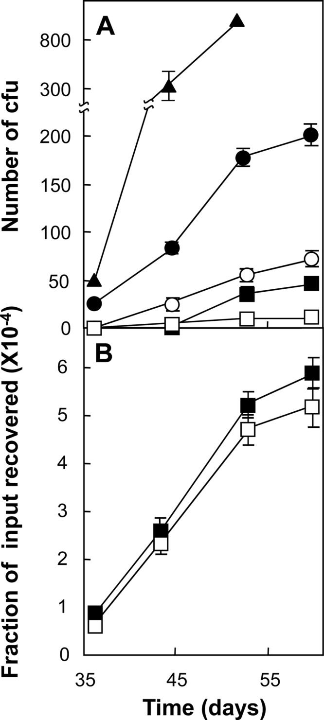
Fig 5.

Ciprofloxacin-resistant mutants of M. tuberculosis induced by ciprofloxacin. (A) Exponentially growing cultures of M. tuberculosis H37Rv (1.5 × 10⁷ CFU) were applied to ciprofloxacin-containing agar and incubated at 37°C. At the indicated times, the number of colonies was determined. Symbols: filled triangles, 0.3 μg/ml (1.5 times the MIC); filled circles, 0.4 μg/ml (2 times the MIC); open circles, 0.5 μg/ml (2.5 times the MIC); filled squares, 0.6 μg/ml (3 times the MIC); open squares, 0.8 μg/ml (4 times the MIC). (B) Induction of ciprofloxacin-resistant mutants by pansusceptible and MDR clinical isolates. Cultures (2.5 × 10⁶ CFU) of a pansusceptible isolate (strain 12850, filled squares) and an MDR isolate (strain 16644, empty squares) were applied to agar containing ciprofloxacin at 2 times the MIC and incubated at 37°C. At the indicated times, the cumulative number of colonies was determined and expressed as a fraction of the number of input cells. Similar results were obtained with two other pairs of pansusceptible and MDR isolates (strains 10775 and 10536 and strains 13571 and 18996). Error bars represent standard deviations; replicate experiments gave similar results.
