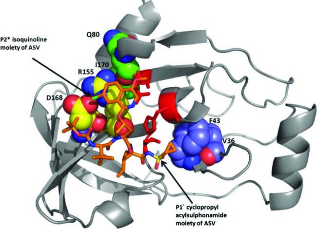
Fig 4.
Model of asunaprevir (ASV) bound to the active site of truncated tether form of the NS3/4A protease (our unpublished data). Asunaprevir is depicted in orange. The NS3 protease is shown as a ribbon diagram. Residues susceptible to changes that confer minimal to high levels of resistance to asunaprevir are depicted using space-filled atoms and labeled.

