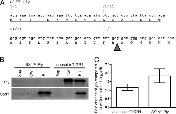
Fig 2.
SSRrgB-Ply fusion mRNA is made but protein is not detected in Streptococcus pneumoniae. (A) N-terminal DNA (top) and amino acid (bottom) sequences of SSRrgB-Ply. Text that is not in bold is Ply sequence. The RrgB signal peptide is in bold and underlined. The arrowhead indicates the predicted site of signal peptidase cleavage. (B) Western blot of cell wall fractionation of SSRrgB-Ply and acapsular TIGR4 cells. Cultures were grown to mid-exponential phase, fractionated into supernatant, cell wall, and protoplast compartments, and assayed for the presence of Ply and CodY by Western blotting. Equal cell equivalents of each fraction were loaded on the gel. A representative Western blot from two independent experiments is shown. Sup, culture supernatant; CW, cell wall; Prt, protoplast. (C) Quantitative RT-PCR on the ply transcript in acapsular TIGR4 and SSRrgB-Ply cells. Shown is the median fold change of the ply transcript of three replicates compared to acapsular TIGR4, normalized to gyrB; error bars indicate the range.

