Fig 3.
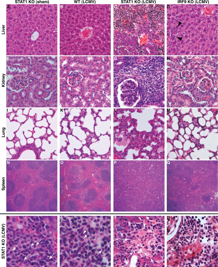
LCMV infection of STAT1 KO mice results in destructive organ pathology. The livers (A), kidneys (E), and lungs (I) of sham-infected STAT1 KO mice showed no overt pathological alterations, and germinal centers present in the spleen appeared normal (N). Comparable findings were obtained for the livers (B), kidneys (F), lungs (K), and spleens (O) from WT mice at day 6 postinfection. In contrast, the livers (C), kidneys (G), and lungs (L) of infected STAT-deficient mice showed prominent mononuclear infiltrates. No organized germinal centers were seen in the spleens (P) of infected STAT1 KO mice. The livers (D, arrowheads), kidneys (H), and lungs (M) from infected IRF9 KO mice contained few foci with infiltrating leukocytes. In the spleens (Q) of infected IRF9 KO mice, germinal centers were less easily discernible than in WT mice but were still identifiable. Higher magnification of tissue sections from the livers (R) and kidneys (S) of infected STAT1 KO mice showed the presence of lymphocytes and polymorphonuclear granulocytes (arrows), as well as focal necrosis, in liver (T, arrows) and kidney (U, arrows; note the normal glomerulus in the top left corner). Original magnifications: A to H, ×20; I to M, ×10; N to Q, ×5; R to U, ×40.

