
Fig 4.
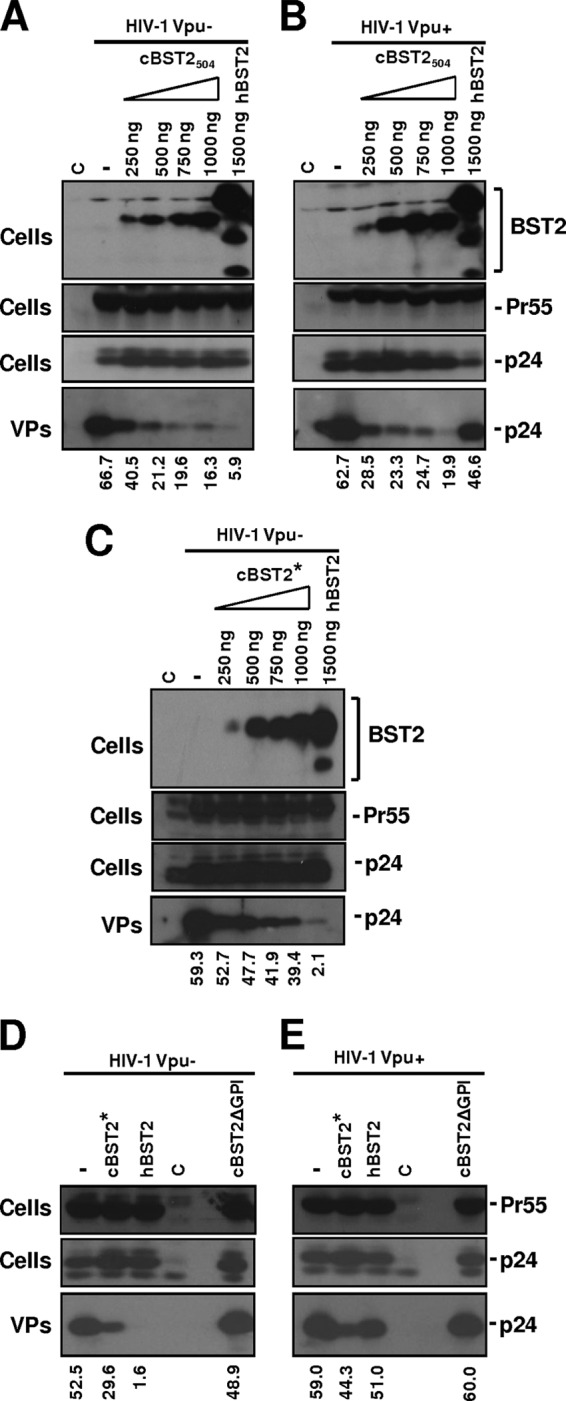
Effect of cBST2 on Vpu− and Vpu+ HIV-1 strain particle release. (A to C) The HIV-1 Vpu−- or HIV-1 Vpu+-expressing constructs (1.25 μg) were transfected into 293T cells alone (−), in the presence of 1,500 ng of hBST2, or along with increasing amounts (250, 500, 750, and 1,000 ng) of cBST2504 (A and B) or cBST2* (C), as indicated. (D and E) 293T cells were transfected with either HIV-1 Vpu−- or HIV-1 Vpu+-expressing plasmids alone (−) or along with 1,500 ng of either the cBST2*, the hBST2, or the cBST2ΔGPI plasmid. At 24 h posttransfection, the cells were lysed, and the viral particles (VPs) were spun down by ultracentrifugation. Proteins derived from the cell lysates (Cells) and from viral particles were analyzed by SDS-PAGE, followed by Western blotting with an anti-Flag monoclonal antibody (BST2) or an anti-HIV-1 Gag monoclonal antibody (Pr55 and p24), as indicated. The numbers below each panel represent the percentage values of particle release, as specified in Materials and Methods. C represents cells transfected with the pcDNA3.1 empty vector.