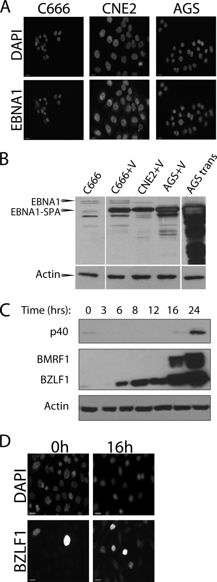
Fig 1.
Expression of SPA-tagged EBNA1 and lytic cycle induction. (A) C666, CNE2, and AGS cells were grown on coverslips and infected with adenovirus to deliver SPA-tagged EBNA1. Forty-eight hours after infection, cells were fixed and stained as previously described (24) using FLAG antibody (Bethyl; 1:800) and goat anti-rabbit Alexa Fluor 555 secondary antibody (Invitrogen; 1:100). Coverslips were mounted onto slides using ProLong Gold antifade medium containing DAPI (4′,6-diamidino-2-phenylindole) (Invitrogen). Images were obtained using the 40× oil objective on a Leica DM IRE2 inverted fluorescence microscope and processed using OpenLAB (version X.0) software. (B) C666, CNE2, and AGS cells were infected with the adenovirus expressing EBNA1-SPA (+V) and, 48 h later, lysed in 9 M urea and 5 mM Tris-HCl (pH 6.8) followed by brief sonication. Similar lysates were also generated of AGS cells 48 h posttransfection with pMZS3F.EBNA1 (22) expressing SPA-tagged EBNA1 (AGS tran) and of C666 cells (first lane). A total of 30 μg of each lysate was analyzed by Western blotting using K67 rabbit serum against EBNA1 (kindly supplied by Jaap Middeldorp). The positions of the native EBNA1 in C666 cells (EBNA1) and EBNA1-SPA are indicated. The latter is smaller than native EBNA1 because it lacks most of the nonessential Gly-Ala repeat region. (C) AGS-EBV cells were treated with 3 mM NaB and 20 ng/ml TPA and harvested 3 to 24 h later as indicated. Cells were lysed in 9 M urea 5 mM Tris-HCl (pH 6.8) and briefly sonicated. A total of 50 μg of total protein was analyzed by Western blotting using antibodies against BZLF1 (Santa Cruz; 1:1,000 dilution), BMRF1 (Chemicon; 1:5,000), VCA-p40 (1:500; kindly supplied by Jaap Middledorp), and actin (Santa Cruz; 1:5,000). (D) AGS-EBV cells grown on coverslips were fixed 16 h after NaB/TPA treatment, stained with antibody against BZLF1 (Santa Cruz; 1:50) followed by anti-mouse Alexa Fluor 488 (Invitrogen; 1:100), and counterstained with DAPI. Images were obtained as described for panel A.

