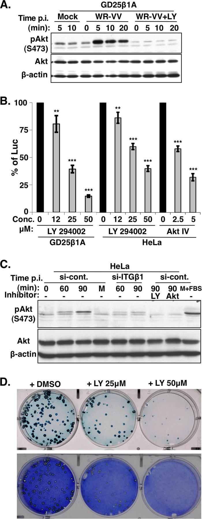
Fig 4.
PI3K/Akt activation induced by WR-VV is required for virus entry. (A) WR-VV induced phosphorylation of Akt in GD25β1A cells. GD25β1A cells were serum starved, pretreated with DMSO or the PI3K inhibitor (LY294002), and either mock infected or infected with medium containing purified MV particles (WR-VV) at an MOI of 60 PFU per cell at 37°C. Cells were harvested at 0, 5, 10, and 20 min after the addition of virus for immunoblot analyses with anti-phospho-Akt (pAkt) (S473), anti-Akt, and anti-β-actin antibodies. (B) Viral early luciferase activity in GD25β1A and HeLa cells was blocked by PI3K/Akt inhibitors. Cells were pretreated with DMSO; the PI3K inhibitor (LY294002) at a concentration of 12, 25, or 50 μM; or the Akt inhibitor (Akt IV) at a concentration of 2.5 or 5 μM and subsequently infected with WR-VV at MOIs of 10 PFU (for GD25β1A cells) and 5 PFU (for HeLa cells) per cell and harvested at 2 h p.i. for luciferase assays as described above. The luciferase activity present in the DMSO-treated samples was used as 100%. The bars represent the standard deviations from three independent experiments. Statistical analyses were performed by using Student's t test in Prism software (GraphPad). P values are shown (**, P < 0.001; ***, P < 0.0001). (C) WR-VV-induced phosphorylation of Akt requires integrin β1 in HeLa cells. The si-control and si-ITGβ1 KD HeLa cells, as described in the legend of Fig. 2A, were serum starved, pretreated with or without inhibitors of PI3K (LY294002) or Akt (Akt IV), and subsequently infected with purified MV particles (WR-VV) at an MOI of 40 PFU per cell at 37°C. Cells were harvested at 0, 60, and 90 min after the addition of virus for immunoblot analyses with anti-phospho-Akt (pAkt) (S473), anti-Akt, and anti-β-actin antibodies. The si-control HeLa cells stimulated with medium containing 20% fetal bovine serum (M+FBS) for 30 min were used as a control. (D) Vaccinia virus plaque formation was reduced with the PI3K inhibitor LY294002 in GD25β1A cells. GD25β1A cells were pretreated with DMSO or the inhibitor LY294002 at concentrations of 25 and 50 μM for 1 h at 37°C and subsequently infected with WR-VV (∼300 PFU/well), cultured in medium containing inhibitors, fixed at 2 days p.i., and stained with X-Gal to visualize the blue plaques. After photography, cells were subsequently stained with crystal violet to reveal the monolayer of cells.

