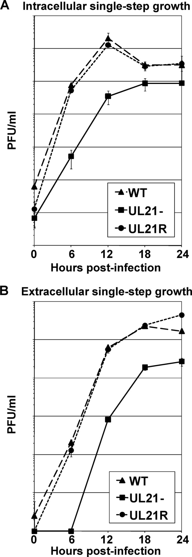
Fig 1.

Single-step growth analyses of the WT, UL21−, and UL21R viruses. Vero cell monolayers were infected with the WT, UL21−, and UL21R viruses at an MOI of 5 PFU/cell for 1 h to allow virus adsorption. The cells were then washed extensively with citrate buffer to neutralize and remove unbound virus. The cells were overlaid with medium and held at 37°C. At the indicated times postinfection, the infected cells (A) and the overlying medium (B) were analyzed separately by plaque assay to determine intracellular and extracellular virus yields, respectively. Data points are the arithmetic means from three independent experiments; error bars represent one standard deviation.
