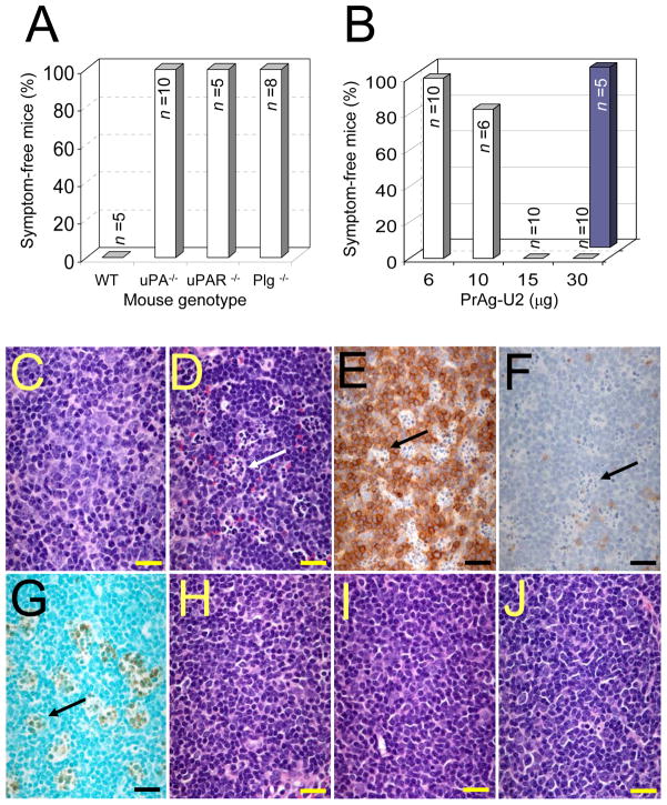
Fig. 3.
uPA-dependent activation of PrAg-U2/FP59 requires the presence of uPA, uPAR, and plasminogen in vivo. (A) Plg, uPA, and uPAR-deficient mice are hyperresistant to uPA-activated anthrax toxin. Wild type mice and mice deficient in uPA, uPAR, and Plg were challenged with 200 μg PrAg-U2 with 10 μg FP59 intraperitoneally, and were monitored for disease. All wild type mice became terminally ill within 24 h of toxin administration, whereas no outwards or histological signs of toxicity were detected in uPA, uPAR, and Plg-deficient mice (P<0.01). (B) PAI-1-deficient mice are hypersensitive to PrAg-U2. PAI-1−/− (open bars) or wild type control (solid bars) mice were challenged with varying concentrations of PrAg-U2 with 10 μg FP59, and monitored for disease. All PAI-1−/− mice treated with 15 to 30 μg PrAg-U2 became terminally ill within 24 h of toxin administration, whereas no outwards or histological signs of toxicity were detected in wild type mice challenged with 30 μg PrAg-U2 (P<0.001). (C–J) Cell-surface uPA-dependent T-cell toxicity of PrAg-U2. Histological appearance of T cell regions of the spleen of wild type (C–G), uPA−/− (H), uPAR−/− (I), and Plg−/− (J) mice 24 h after intraperitoneal injection of PBS (C) or 200 μg PrAg-U2 with 10 μg FP59 (D–J). Scattered clusters (examples indicated with arrows) of degenerating lymphocytes in wild type mice (D), absent in PBS-treated wild type mice (C), are identified as subpopulations of T-cells, by immunostaining with T-cell (E) and B-cell (F) antibodies, undergoing apoptosis as visualized by TUNEL-staining (G). (H–J) shows the absence of T-cell pathology in the spleens of uPA−/− (H), uPAR−/− (I) and Plg−/− (J) mice. (C, D, and H–J) Hematoxylin/eosin staining. (Bars = 10 μm).

