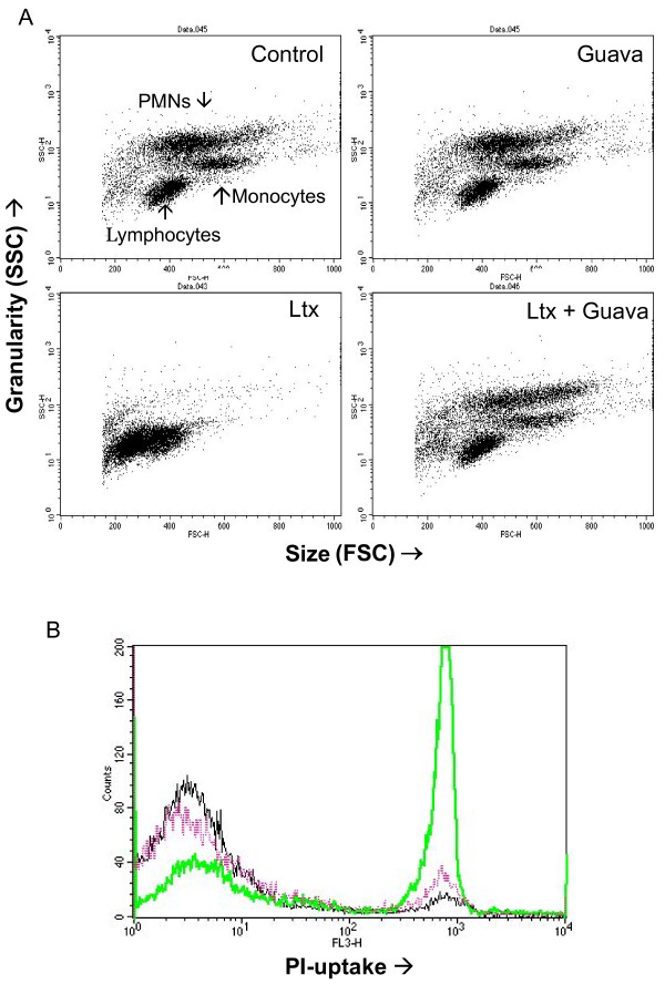
Figure 3.
Leukotoxin-induced morphological alterations in monocytes and neutrophils are abolished in the presence of Guava extract. Human leukocytes exposed to A. actinomycetemcomitans leukotoxin (100 ng/ml) for 60 min with or without extracts (0.1%) from Guava twigs. Cell morphology of the different exposed leukocyte subsets (FSC/SSC) was documented by flow cytometry analyses (A). Viability (PI-uptake) in the exposed leukocyte populations was quantified by flow cytometry analyses (B). Black line represents control cells, green line leukotoxin-exposed cells and the violet line cells exposed to leukotoxin in the presence of Guava extract. Representative dot plots and histogram are shown.

