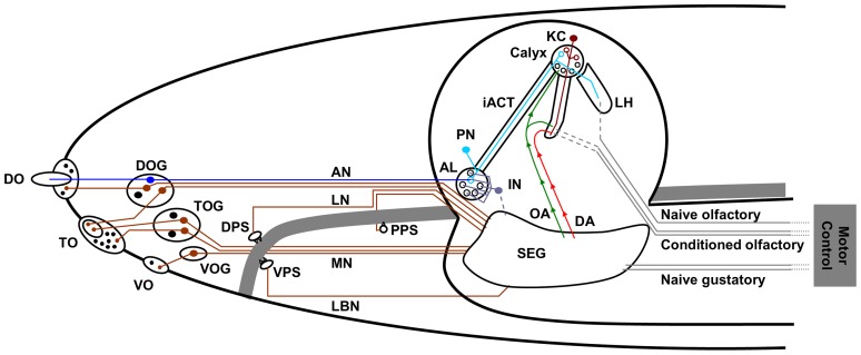
Figure 1. The anatomy of the Drosophila chemosensory system.
Overview of the cephalic larval chemosensory pathways. Olfactory pathways (blue) project into the brain proper, whereas gustatory afferents (brown) are collected in various regions of the subesophageal ganglion. The green and red arrows indicate pathways to short-circuit a taste-driven reinforcement signal from the subesophageal ganglion towards the brain. Note that the different gustatory organs project to different regions in the subesophageal ganglion. Abbreviations: AL: antennal lobe; AN: antennal nerve; DA: dopaminergic neurons as engaged in reinforcement signalling; DO/ DOG: dorsal organ/ dorsal organ ganglion; DPS: dorsal pharyngeal sensillae; iACT: inner antenno-cerebral tract; IN: antennal lobe interneurons; KC: Kenyon cells; LBN: labial nerve; LH: lateral horn; LN: labral nerve; MN: maxillary nerve; OA: octopaminergic neurons as engaged in reinforcement signalling; PN: projection neurons; PPS: posterior pharyngeal sensillae; SEG: subesophageal ganglion; TO/ TOG: terminal organ/ terminal organ ganglion; VO/ VOG: ventral organ/ganglion; VPS: ventral pharyngeal sensillae. Based on Stocker 2008 [10].

