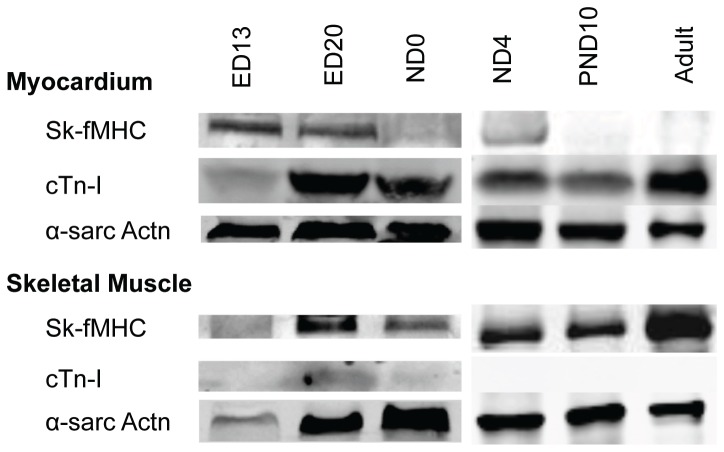
Figure 3. Western blot analysis of developing myocardium and skeletal muscle.
Lane 1: ED13; Lane 2: ED20; Lane 3: ND0; Lane 4: ND4; Lane 5: PND10; Lane 6: Adult (8 week-old). Top panel: Left ventricular myocardium, Bottom panel: hind limbs at ED13, ED20, ND0, and ND4, and gastrocnemius muscle in PND10 and adult. At ED13 and ED20, left ventricular myocardium expressed sk-fMHC and cTn-I expression was very weak while skeletal muscle expressed sk-fMHC. During development, sk-fMHC expression in the left ventricular myocardium decreased and increased in skeletal muscle, whereas cTn-I is expressed in the left ventricular myocardium and was negative in skeletal muscle. α-sarcomeric actinin (α-sarcActn) was used to adjust the total protein loading for the electrophoresis.

