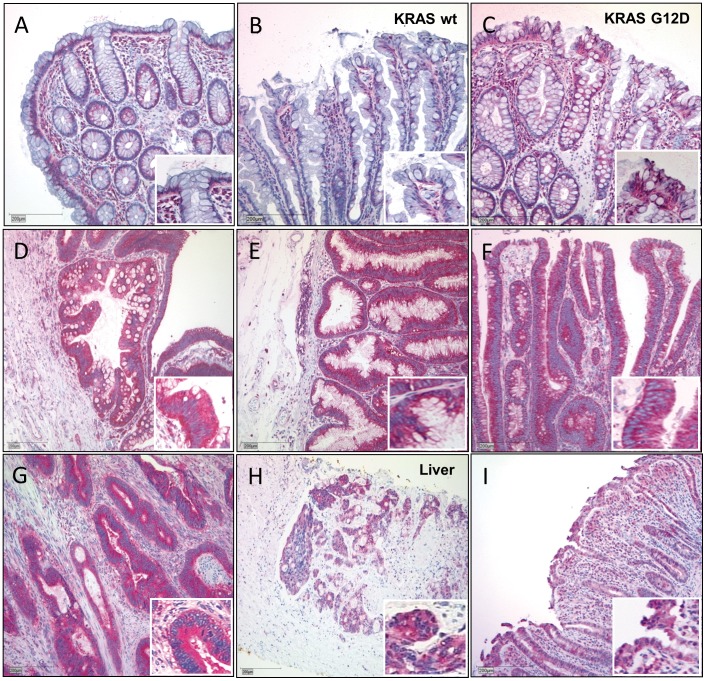
Figure 1. Abi1 immunohistochemistry in colorectal tissue samples. A.
and B, Regular mucosa (A) and wild-type hyperplastic polyps (B) show only moderate, cytoplasmatic and basally located Abi1 immunoreactivity. Note intense staining of lymphocytes in subjacent stroma. C, Overexpression of Abi1 in a representative hyperplastic polyp harboring KRAS G12D mutation. D-F, Intense immunoreactivity in (wild-type) sessile serrated adenoma (D), traditional serrated adenoma (E) and tubular adenoma (F). Note that positivity for Abi1 is exclusively cytoplasmic. G and H, strong immunoreactivity for Abi1 in invasive colorectal carcinoma and in a liver metastasis of colorectal carcinoma. I, strong cytoplasmic immunoreactivity for Abi1 in inflamed colonic mucosa. Stain: anti-Abi1, haematoxylin; Bar indicates 200 µm.

