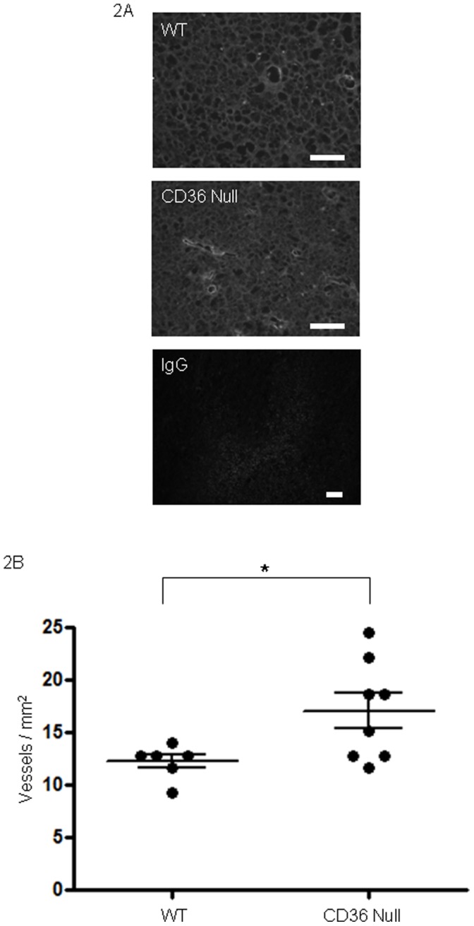
Figure 2. Cd36 deletion in mice enhances Lewis Lung tumor vascularity.

(A) Lewis Lung tumors as in Figures 1 were dissected, sectioned and examined by immunofluorescence microscopy using anti-VEGF receptor antibody (green) to detect blood vessels. DAPI stained nuclei are blue. Magnification bars represent 100 µm. IgG control is shown in bottom panel as negative control. (B) Vessel densities measured as vessels per mm2. Median vessel density: wt 12.83, cd36 null 16.91.
