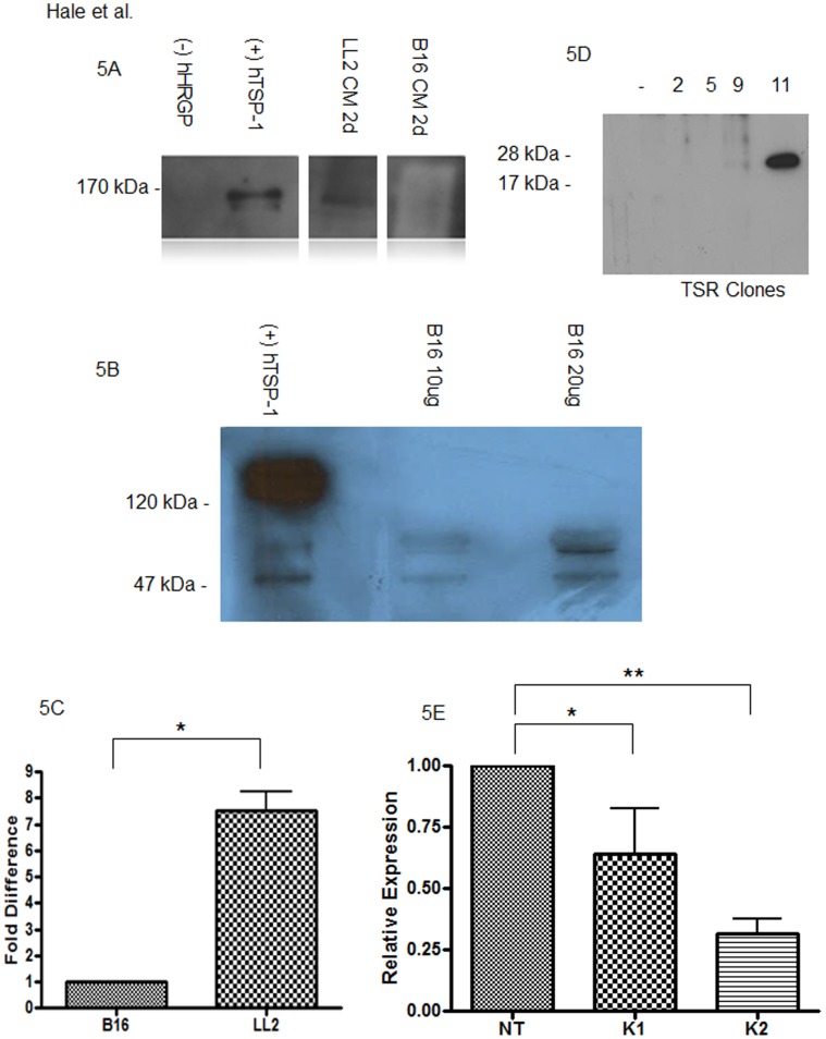
Figure 5. Thrombospondin-1 expression in LL2 and B16F1 melanoma cells and tumors.
(A) Lewis Lung (LL2) or B16F melanoma cells were cultured in serum free media for 24 hours (1d) at which point proteins in post culture media (CM) were precipitated by TCA, separated under reducing conditions by SDS/PAGE and analyzed by immunoblot using anti-TSP-1 antibody. TSP-1 monomers were detected at 170 kDa in the media conditioned by LL2 cells, but not B16F1 cells. Purified human HRG and TSP were used as controls. (B) B16F1 melanoma tumor tissue was analyzed by western blot analysis for TSP expression. Intact TSP was not observed at 150 kDa, however possible degredation products were observed around 55 kDa. (C) B16F1 and LL2 tumor tissue was analyzed by RT-PCR for expression of TSP. TSP was detected in both tumor types, approximately 7 fold higher in LL2. (D) Conditioned media was collected from 4 different antibiotic resistant clones of TSR transfected B16F melanoma cells and analyzed by immunoblot as in panel A. Clone 11 expressed abundant anti-TSP reactive material at the appropriate molecular weight of recombinant TSR and was utilized for subsequent tumor studies. (E) TSP knockdown efficiency was analyzed by RT-PCR with statistical significance as indicated; **P<0.05; *P = 0.06. In both instances of TSP knockdown 1 (K1 and K2), reductions in TSP message levels were detected as compared with nontargeted control (NT) cells.

