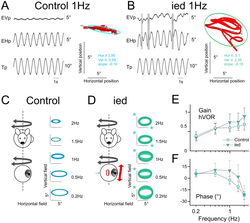
Figure 1. Horizontal angular vestibulo-ocular reflex in C57Bl/6J and ied mice.
A–B, Example of eye movements evoked in C57Bl/6J (A) or ied (B) mice during 1 Hz sinusoidal oscillation in horizontal. Shaded areas indicate quick phases. Plots present oculomotor fields. Red points are the eye position of the same traces. The ellipses present 95% of the horizontal and vertical eye positions. θ are horizontal and vertical variance; inclination of the ellipse was computed as the slope of the linear regression between vertical and horizontal eye positions. C–D Averaged oculomotor fields in C57Bl/6J (C) and ied (D) mice across tested frequencies. Line and surface of the ellipses present the mean and standard deviation of the population, respectively. The slopes of the ellipses are the mean slope of the individuals’ ellipses. Green asterisks indicate significantly larger response in ied compared to C57Bl/6J. E–F, Gain (E) and timing (F) of the horizontal component of eye movement responses for C57Bl/6J and ied populations. Asterisk indicates statistical difference with p<0.05. Tp, Table position; EVp, Eye Vertical position; EHp, Eye Horizontal position. In this and following figures, plots present mean ± SD.

