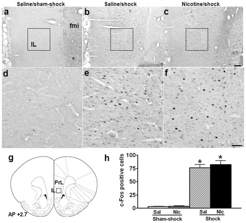
Fig. 3.
Effect of nicotine self-administration on stress-induced neuronal activation in infralimbic cortex. Photomicrographs (a–c) illustrate immunohistochemical labeling of c-Fos in coronal sections of infralimbic cortex (IL). Boxes indicate the positions of areas used for counting c-Fos+ neurons; these areas are shown in panels d-f at higher-magnification. A schematic representation of IL is shown in panel g, with the position of the counting square (380 × 380 μm2) shown. Both saline (a, d) and nicotine (images not shown) rats expressed c-Fos at very low levels after sham-shock. Mild footshock stress increased c-Fos expression to comparable levels in both the saline (b, e) and nicotine (c, f) groups. The mean ± SEM number of stress-induced c-Fos+ neurons is shown in panel h. *: p < 0.01, compared to the corresponding sham-shock controls. Scale bars: panels a–c, 100 μm; panels d–f, 50 μm. fmi: forceps minor of the corpus callosum.

