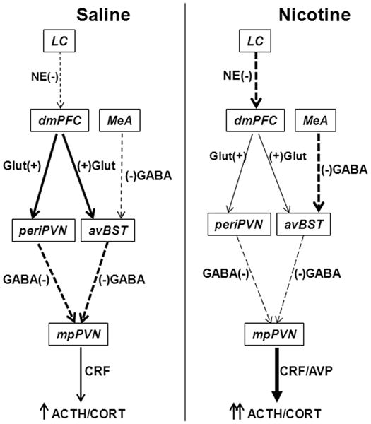
Fig. 8.

Schematic diagram of the effects of chronic nicotine self-administration on stress-induced neuroactivity in the limbic-PVN network. Nicotine diminished stress-induced activity of glutamatergic (Glut) excitatory (+) neurons (designated by thinner solid arrows compared to saline control) that project from prelimbic cortex (PrL) to anteroventral bed nucleus of the stria terminalis (avBST) and to peri-PVN. The reduced activity of PrL neurons may result from the enhanced activity of norepinephrinergic (NE) inhibitory (−) neurons (thicker dashed arrow compared to saline) projecting from locus coeruleus (LC). In contrast, nicotine increased the activity of GABAergic inhibitory (−) neurons that project from medial amygdala (MeA) to avBST. Therefore, the activities of GABAergic inhibitory (−) neurons projecting from avBST and peri-PVN to medial parvocellular PVN (mpPVN) were diminished. These effects of chronic nicotine on the limbic-PVN network result in the disinhibition of corticotrophin-releasing factor (CRF) neurons by reduced stress-induced GABA release. This disinhibition facilitates the stimulation of CRF neurons and the release of CRF and arginine vasopressin (AVP) by the enhanced stress-induced PVN glutamate release, which we have previously reported (Yu et al. 2008, Yu et al. 2010).
