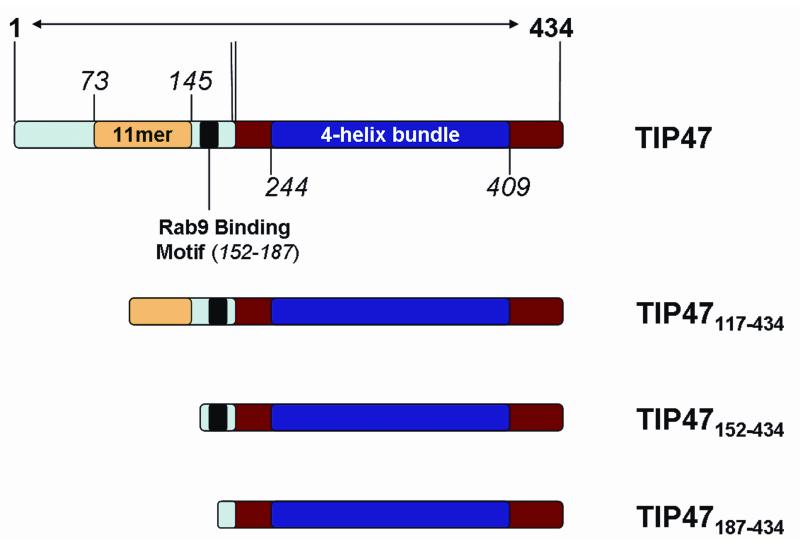
Figure 1.
Domain structure of full-length human TIP47/perilipin-3 and the N-terminal constructs generated for this study. The N-terminal regions are highlighted in light blue with yellow and black inserts indicating the 11-mer repeat and Rab9 binding regions, respectively. The C-terminal region corresponding to the mouse crystal structure is in red, with the 4-bundle helix region as a dark blue insert.

