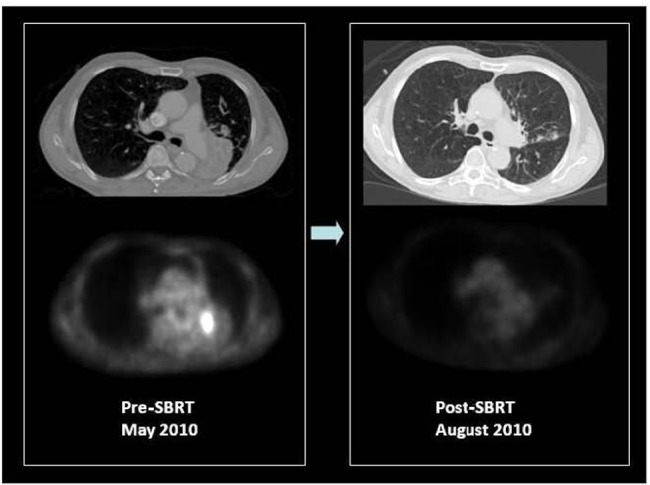
FIGURE 4.

F18 FDG PET/CT of a patient with medically inoperable, centrally located, stage I squamous cell carcinoma of the left lower lobe causing obstructive left lower lung collapse. Contrast CT is unable to differentiate tumor from adjacent collapsed left lower lobe. PET only image of the same axial slice demonstrates FDG avidity of only the tumor and differentiates metabolically active tumor from adjacent collapsed lung. PET was used for stereotactic body radiation therapy image-guided treatment planning to target the FDG avid tumor. Patient received 50 Gy in four fractions in May 2010. Early post-SBRT PET/CT imaging was performed in August 2010 to assess therapy response, which showed a complete metabolic response and re-inflation of the left lower lobe. The patient is currently free of local or distant recurrence or post-radiation complications with improved exercise tolerance and lowered oxygen consumption. Courtesy of UCLA Radiation Oncology SBRT program.
