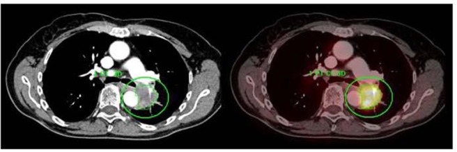
FIGURE 7.

A centrally located left upper lobe tumor. F18 FDG PET/CT shows large FDG avid mass without mediastinal metastases. Integrated contrast CT defines the relationship of tumor with adjacent aorta and pulmonary vessels. Automated 3D contouring of metabolically avid tumor was performed using a threshold of 40% of SUVmax. Volume of interest was exported as RT structure set for radiation planning.
