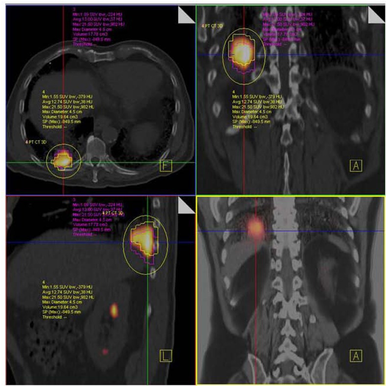
FIGURE 9.
NSCLC at right lung base. F18 FDG PET/CT shows hypermetabolic tumor mass close to diaphragm and posterior thoracic wall without nodal or distant metastases. Respiratory-gated PET/CT was performed. Images show end inspiratory (pink) and end expiratory (yellow) metabolic tumor volumes superimposed on non-gated PET/CT image demonstrating tumor motion during the entire respiratory cycle.

