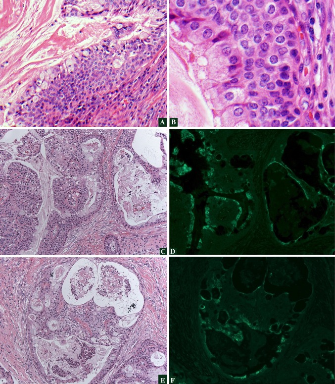
Fig. 1.
HPV and Histologya, b demonstrate low- and high-power magnification, respectively, of an HPV-positive, cystic, low-grade MEC with proliferation of basaloid type cells. Immunofluorescence (IF) for HPV16/18 E6 protein in MECc, e demonstrate hematoxylin and eosin stained areas of a MEC which is HPV-positive. d, f represent the corresponding regions demonstrating positive IF staining using an antibody to HPV16/18 E6 protein. Tumor nuclear and cytoplasmic staining is seen (bright green) which correlates with both the glandular and squamoid elements

