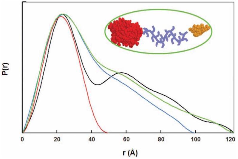
Fig. (1).
Experimental P(r) functions of multidomain proteins. Experimental P(r) functions of the Humicola insolens cellulase Cel45 and variants: globular catalytic domain (red curve), catalytic domain and linker (blue curve), full-length Cel45 wild-type (green curve), and full-length Cel45 with a proline mutation leading to a more rigid linker (black curve). The crystal structures of the catalytic domain (red) and the cellulose-binding domain (yellow) are represented in space-filling mode. The enhanced rigidity of the linker in the mutant Cel45 translates into a P(r) function with a well separated peak corresponding to the interdomain distances. (Figure adapted from [60]).

