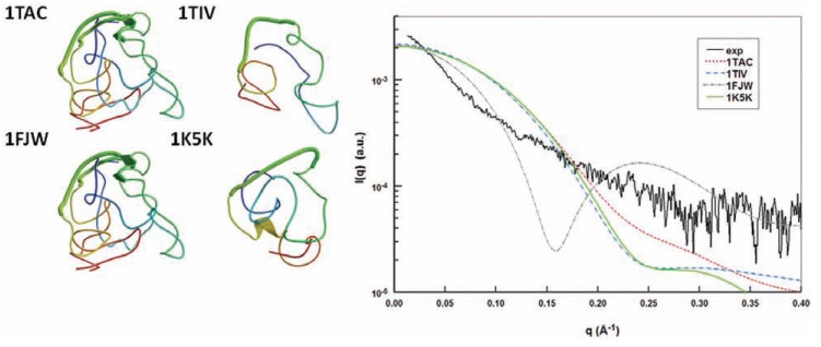
Fig. (6).
Use of CRYSOL to compare atomic structure with the structure in solution observed by SAXS. Comparison of experimental SAXS data from HIV-TAT (black line) with the theoretical scattering curve of published structures of TAT with pdb code 1TAC (red dotted line), 1TIV (blue dashed line), 1FJW (grey dash-point line), and 1K5K (continue green line)] using the CRYSOL program to show the discrepancy between the pdb structures and the structure in solution observed by SAXS. The low statistics of the experimental curve are accounted for by the low concentration of the protein (< 1 mg/mL) (Figure adapted from [102]).

