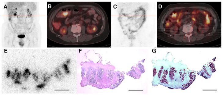
FIGURE 2.
(A–B) 18F-FDG PET/CT scan acquired immediately before administration of 124I-huA33 shows pathologic uptake in lesion in transverse colon near hepatic flexure together with normal physiologic accumulation. (C–D) PET/CT scan acquired 182 h after administration of 140 MBq (3.8 mCi) of 124I-huA33 shows focal uptake in tumor (compare B and D) and throughout normal intestine. (E–G) Contiguously aligned sections of tumor visualized in D assessed by 124I-DAR (E), hematoxylin and eosin staining (F), and immunohistochemical staining (G) for A33 antigen. 124I-huA33 localized only in small (~1 mm in dimension) regions of viable antigen-positive tumor and not in antigen-negative stroma. Scale bars = 2 mm.

