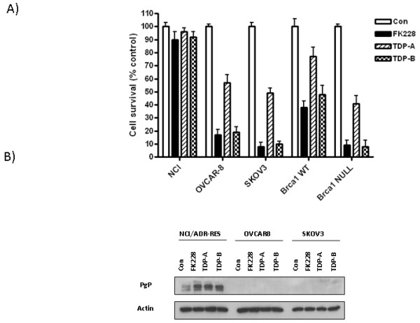
Figure 2.

TDP-A and TDP-B inhibit ovarian cancer cell survival at nanomolar concentrations. A) Representative graphs of NCI/ADR-RES, OVCAR-8, SKOV-3, BRCA1 wild type and null ovarian cancer cells treated with FK228, TDP-A and TDP-B at a fixed concentration of 10 nM. MTT assays were performed to assess cell proliferation after 72 h of treatment. Each treatment was replicated 6 times. Values are mean + SE for 3 independent experiments. B) Representative Western blot for P-glycoprotein expression in NCI/ADR-RES, OVCAR-8 and SKOV-3 ovarian cancer cells after 24 h of treatment with 10nM FK228, 10nM TDP-A and 10nM TDP-B. 0.1% DMSO was the vehicle control. β-actin was used as a loading control.
