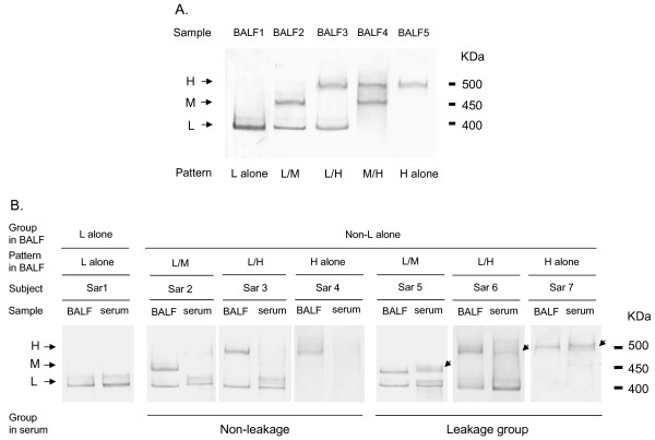
Figure 2.
A. Representative examples of Western blot analyses using anti-KL-6 antibody in BALF. Three bands were detected under reducing conditions with low (L), middle (M) and high molecular weights (H), corresponding to approximately 400, 450 and 500 kDa, respectively. Based on the three bands, there were five band patterns (L alone, L/M, L/H, M/H, H alone) identified in 128 subjects with sarcoidosis. B. Comparison of KL-6/MUC1 band patterns between BALF and serum in 7 representative subjects. Sar, subjects with sarcoidosis. Arrows indicate M or H bands present in serum.

