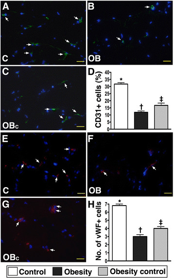
Figure 5.
Quantifications of endothelial cells (ECs) in ischemic region by day 14 after CLI procedure using immunofluorescent microscope (400x) (n = 9). A to C) showing the numbers of CD31+ ECs (white arrows) remarkably higher in normal control (C) group than in obesity (OB) and obesity control (OBC) groups, and significantly higher in OBC group than in OB group. D) * vs. other groups with different symbols, p < 0.0001. E to G) showing the numbers of von Willebrand factor (vWF) + ECs (white arrows) notably higher in C group than in OB and OBC groups, and significantly higher in OBC group than in OB group. G) * vs. other groups with different symbols, p < 0.0001. Statistical analysis by ANOVA followed by Bonferroni multiple comparison post hoc test. The scale bars in right lower corner represent 20 μm.

